
The IOLMaster 700
Better predictability and optimized workflow compared with other biometers.
BY OLIVER FINDL, MD, MBA, FEBO
ntroduction of the first Swept Source OCT-based biometer, the IOLMaster 700 (Carl Zeiss Meditec; Figure 1) enables OCT imaging and visualization across the entire length of the eye. In comparison to A-scan biometry, in which the eye is visualized in one dimension only, the IOLMaster 700 provides an image-based measurement, allowing one to view the complete longitudinal section of the eye—from the cornea to the retina.
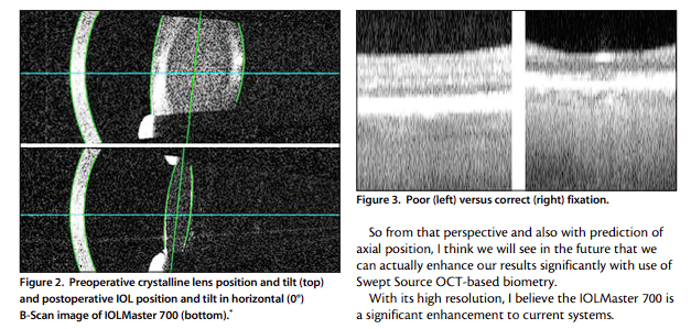
Therefore, the IOLMaster 700 can also be used to identify irregular eye geometries, such as lens tilt (Figure 2), and, thanks to imaging of the fovea in the macula, to alert the user to insufficient fixation during measurements (Figure 3). Furthermore, use of telecentric keratometry allows particularly robust and reproducible corneal surface measurements. For these reasons and more, the IOLMaster 700 is an advantageous diagnostics device for the anterior segment surgeon in that it helps to significantly improve refractive results after cataract surgery.
HIGH PRECISION, SIMPLIFIED WORKFLOW
Because OCT images can be captured along the entire length of the eye, the IOLMaster 700 allows identification of all ocular peaks and interfaces. This helps in selecting the best measurements, not only for axial length but also for keratometry (K). This is especially important in patients who do not always fixate on the light. Off-axis fixation during keratometry also results in false K readings. The absence of the foveal pit in the IOLMaster 700’s retinal scan can be an indication for off-axis fixation. By eliminating all measurements in which the patient was not fixated, I have even better and more precise measurements than before. As with the IOLMaster 500, the latest version simplifies my preoperative workflow. In eyes with astigmatism, the
IOLMaster 700 acquires
a reference image of the scleral and conjunctival vessels. The image of the eye is taken along with the keratometry measurement and directly transferred, over the hospital’s intranet, to the CALLISTO eye (Carl Zeiss Meditec) computer in the operating theater. CALLISTO eye is connected to the LUMERA microscope (Carl Zeiss Meditec). During surgery, the image allows intraoperative matching with the live eye image, eliminating the need for manual pre- and intraoperative markings of the astigmatism axis in conjunction with implantation of a toric IOL. It also avoids manual data transfer because the images are directly transferred over the hospital’s intranet. There is also a built-in toric IOL calculator in the IOLMaster 700. Other advantages of the IOLMaster 700 are its compatibility with previous versions and that it provides access to the database of the User Group of Laser Interference Biometry (ULIB), containing the lens constants of more than 270 IOL models.
PATIENT EXPECTATIONS
Patient education is important. With rising demands and expectations, patients want to know not only what is happening, but also what the surgeon is doing. With other biometry systems, the surgeon was only able to show patients some peaks, and that was not only abstract for the patient but also difficult to understand. With the IOLMaster 700, patients can view a scan of the entire eye—with the cornea, the lens, the retina, and the pupillary edge. This helps patients to understand what the surgeon sees and what he or she will try to accomplish with surgery. It is easier to tell patients where the artificial lens will be positioned and, if they have a tilted lens, to explain that there is a possibility that the IOL may also be tilted. Patients are fascinated by images, and the IOLMaster 700 gives us the capability to appeal to this side of their intellect.
ENHANCING REFRACTIVE OUTCOMES
In my short-term experience with the IOLMaster 700, I have seen better refractive outcomes as a result of the highly precise measurements. In the future, this device may allow us to be much more precise in our predictions of effective lens position and in our imaging of the posterior corneal surface. Swept Source OCT with the IOLMaster 700 will open many doors with regard to biometry and IOL power calculation.
IOL POSITION
Effective IOL position is still the holy grail of IOL power calculation and its prediction error is the main source of refractive surprises in modern cataract surgery. However, now that we can actually see the entire lens— the front and back surfaces and not just peaks—we at least can see a tilted, and maybe even decentered, lens already. In future, we might be even able to consider tilt and decentration in IOL power calculation; however, more data is needed to confirm that.
So from that perspective and also with prediction of axial position, I think we will see in the future that we can actually enhance our results significantly with use of Swept Source OCT-based biometry. With its high resolution, I believe the IOLMaster 700 is a significant enhancement to current systems.
CONCLUSION
The use of Swept Source OCT in biometry, whereby the entire eye is scanned three dimensionally, allows me to not only better understand the eye before surgery but also to have better refractive outcomes after surgery. The technology is now here, and we have many things to learn in the coming months and years with regard to what the IOLMaster 700 can do.


